MITRAL STENOSIS:
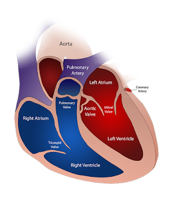
The Mitral valve is one of the four valves present in the human heart. The Mitral valve is situated between the left atrium and left ventricle. It functions by opening during diastole which helps in the forward movement of oxygenated blood from the left atrium into the left ventricle and during ventricular systole this valve closes to prevent backflow of blood from the left ventricle back into the left atrium.
The components of the mitral valve are the annulus, chordae, leaflets, and papillary muscle. The Mitral valve has two leaflets and these leaflets are attached to the annulus which helps to maintain the shape of the valve.
Proper functioning of the mitral valve depends on these components and damage to any of these components can lead to improper functioning of the mitral valve.
The mitral valve’s normal opening is about 4-6 cm2 and a fixed amount of blood passes from the left atrium to the left ventricle through it.
MITRAL STENOSIS: It is a condition in which the normal opening area of the valve is reduced and the blood passage from the left atrium to the left ventricle gets reduced.
It is more common in females as compared to males.
CAUSES OF MITRAL STENOSIS:
1) RHEUMATIC HEART DISEASE: Most common cause of mitral stenosis.
Some less common causes include:
2) CALCIFICATION OF ANNULUS: seen in older patients mostly.
3) CONGENITAL MITRAL STENOSIS
4) CARCINOMA OF THE LUNGS.
5) INFECTIVE ENDOCARDITIS
PATHOPHYSIOLOGY:
As we know that the mitral valve acts as a connection between the left atrium and the left ventricle, so when the opening of the valve is stenosed or reduced in size then it produces resistance to the blood flow from the atrium to the ventricle.
Due to this less blood passes into the ventricle and more blood is accumulated in the left atrium, which leads to an increase in the size of the left atrium known as LEFT ATRIAL DILATATION.
Patients with left atrial dilatation are at high risk of developing ATRIAL FIBRILLATION AND CLOT FORMATION.
Due to mitral stenosis, the pressure inside the left atrium increases, and this leads to an increase in the pressure in the pulmonary vein and hence the pressure in the pulmonary artery also increases which leads to PULMONARY HYPERTENSION.
When the pressure in the left atrium increases and if it is not managed in time and adequately then it slowly starts to damage the right atrium and ventricle too.
SYMPTOMS:
1) During early mitral stenosis patient may remain asymptomatic.
2) With the progression of stenosis patient can complain of breathlessness while walking or doing any work.
3) Easily getting fatigued while walking or doing work.
4) Swelling in legs or whole body.
5) Feeling breathlessness while sleeping at night.
6) In severe cases patient can get breathless while eating too which can lead to complaints of weight loss by the patient.
7) Chest infection like cough is common.
SIGNS:
1) MALAR FLUSH: The patient can present with reddish color cheeks.
2) Prominent “ a “ wave of jugular venous pressure can be noted if the patient has high pulmonary pressure.
3) Murmur can be heard by the doctor on auscultation of the patient.
INVESTIGATION:
1) COMPLETE BLOOD COUNT: It can reveal any infection present in the body.
2) D-DIMER: This blood test can reveal the chances of the formation of blood clots in the body.
3) INR ( INTERNATIONAL NORMALIZED RATIO ) should also be monitored.
4) X-RAY: Can show an increase in the size of the left atrium and KERLY B lines which are horizontal lines seen at the base of the lungs near the pleura can also be seen.
5) ECG: Left atrium enlargement can be seen on ECG as NOTCHED P WAVE which is known as P MITRALE.
6) ECHOCARDIOGRAPHY: One of the most reliable investigations for the diagnosis of mitral stenosis as it reveals the amount of stenosis of the mitral valve, the size of the four chambers of the heart, and the degree to which they are affected due to stenosis. The presence of any clots can also be seen.
MANAGEMENT:
1) As rheumatic fever is considered the most common cause of mitral stenosis so the doctor gives appropriate antibiotic treatment to prevent any further damage to the valve.
2) If edema is present then the doctor gives diuretics to prevent the accumulation of fluid in the body.
3) Antiarrhythmic medicines are given if an arrhythmia is diagnosed.
4) Anticoagulation therapy is given by the doctor to prevent clot formation if clot formation is suspected.
5) Surgical intervention such as mitral Valvuloplasty, valvotomy, or valve repair is the mainstay of the treatment.
PREVENTIVE MEASURE:
If a person is suffering from mitral stenosis then the patient should :
1) Stop smoking and drinking alcohol.
2) Avoid eating fatty and oily food.
3) Reduce salt intake.
4) Take medicine properly as prescribed by the doctor.
5) Avoid lifting heavyweight.
Early diagnosis and treatment of mitral stenosis are important to prevent the patient from developing serious complications and it’s important to follow doctor advice.


