PERICARDIAL EFFUSION –
The pericardium is the outer covering of the heart and the space between the heart and the pericardium is known as the pericardial sac. This space is usually filled with very little liquid, but due to some disease conditions, this space can get filled with excessive liquid, known as pericardial effusion.
CAUSES –
1) IDIOPATHIC
2) TUBERCULOSIS
3) HEART FAILURE
4) MYOCARDIAL INFARCTION
5) VIRAL INFECTION
6) TRAUMA
7) HYPOTHYROIDISM
8) AORTIC DISSECTION
9) CANCER ( most commonly lung cancer)
10) Medicine like isoniazid, hydralazine, procainamide.
PATHOLOGY-
Pericardial effusion can occur in two ways:
The first type is when the accumulation of fluid occurs at a rapid pace which leads to tamponade. It mostly occurs in trauma or aortic dissection and mostly blood is filled in the sac. Tamponade causes compression of the heart and impaired filling of the heart chamber. It is a serious condition and requires immediate medical attention to save the life of the patient.
In the second type, the accumulation of fluid occurs at a slow pace and it slowly stretches the pericardium and mostly does not cause tamponade.
CLINICAL FEATURES-
In the case of tamponade-
1) The patient can complain of difficulty in breathing and difficulty increases when the patient gets in the lying down position.
2) The patient can be worried or nervous in appearance.
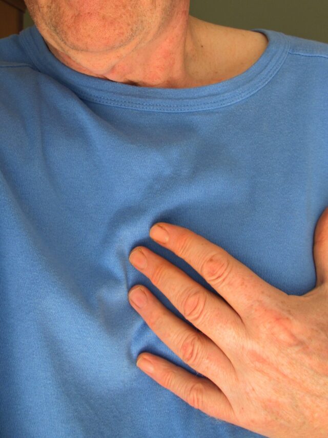
3) Chest pain can be present too.
4) Swelling of the limbs can be seen due to heart failure.
5) Pain in the right side of the stomach which is mostly due to the liver as the tamponade leads to heart failure which in turn causes liver congestion.
In case of slowly developing pericardial effusion-
The patient usually remains asymptomatic and effusion can be noticed with the investigation.
INVESTIGATION-
1) Thyroid function test, diabetes, liver function test, and kidney function test should be done as uncontrolled levels of these are seen in cardiac tamponade.
2) Paradoxical pulse is seen in tamponade patients in which the inspiratory systolic pressure increases by more than 10 mm of Hg.
3) Heartbeat sound will be decreased on auscultation.
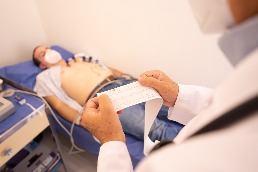
4) ECG will show low voltage.
5) Echocardiography is a very important test to perform if the patient is suspected of pericardial effusion.
6) Chest X-ray shows cardiomegaly.
7) CT and MRI are also important diagnostic tools.
8) Tuberculin skin test, complete blood count, and platelet count should be done to rule out any infections, particularly tuberculosis as these patients are known to develop pericardial effusion at later stages of their life.
TREATMENT-
1) PERICARDIAL EFFUSION WITHOUT TAMPONADE-
In the case where no tamponade is suspected, then it is better to keep the patient under observation and give treatment according to symptoms.
After the blood report and other investigation results come then we should treat the underlying cause of pericardial effusion to improve the patient’s health.
2) CARDIAC TAMPONADE-
a) Drainage of the effusion is important and should be done on an urgent basis.
b) Mechanical ventilation can cause a fall in blood pressure as positive intrathoracic pressure decreases cardiac filling.
c) Echocardiography-guided percutaneous pericardiocentesis can be performed if the patient is in serious condition.
TAKE HOME MESSAGE-
Recurrent pericardial effusion can cause weight loss, fatigue, and breathlessness.
Always take advice from your doctor if you have any of the symptoms or diseases mentioned above.
If you are suffering from hypothyroidism, diabetes, or tuberculosis then it is important that you take your medicine regularly to prevent this disease from happening.
Medicine should be taken on the doctor’s advice as some medicines can increase the risk of pericardial effusion.
After any chest injury, it is important to do chest a X-ray to rule out any serious chest trauma
STAY HEALTHY AND SAFE.


