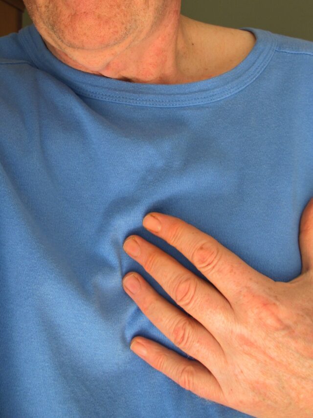
BROKEN HEART SYNDROME CARDIOMYOPATHY-
Broken heart syndrome is a type of cardiomyopathy that is also known as TAKOTSUBO CARDIOMYOPATHY.
Broken Heart syndrome-
Broken Heart Syndrome is an acute stress-induced cardiomyopathy that occurs in typically old age women mostly after emotional or physical stress.

Broken heart syndrome symptoms-
It presents with symptoms that include
- Pulmonary edema presents as breathing difficulties, tightness in the chest, and coughing.
- Hypotension can make the patient Dizzy, weak, and easily fatigued.
- Chest pain.
Investigation-
Cardiac MRI shows diffuse myocardial edema without necrosis of the cardiac cells.
Coronary angiography may be needed to check for acute coronary occlusion.
The ECG may show ST-segment elevation, especially in leads V3 and V4. QT wave prolongation can also be seen.
Management-
- No specific treatment is present for this disease.
- Patients are treated according to the symptoms they present with such as nitrates are given for pulmonary edema.
- If the patient is hemodynamically stable then alpha and beta blockers are given combined.
- Magnesium is also given to the patients if arrhythmia is present.
CARDIOMYOPATHY-
Cardiomyopathy is made up of the terms Cardio, which means cardiac or heart, myo, which means muscle, and pathy, which means disease. So, the meaning of this term is the disease of cardiac muscle. It should be used only in diseases that directly affect the heart muscle and not in diseases whose secondary effects lead to changes in heart muscle such as ischemia, hypertension, and pericardial disease.
CLASSIFICATION OF CARDIOMYOPATHY:
Primary cardiomyopathy-
1) DILATED– Dilation can be seen in the left, right, or both ventricles of the heart. When the ventricle is dilated then it can lead to improper contraction of the ventricle. This causes pooling of blood in the ventricle and cardiac failure.
2) RESTRICTIVE – It can occur due to scarring or infiltration in the cardiac muscle which leads to restriction in movement of these muscles and hence causes improper filling of the blood in the ventricle.
3) HYPERTROPHIC – It is mostly seen in the interventricular septum (septum present between the right and left ventricle of the heart) and also in the left ventricle.
Secondary cardiomyopathy-
1) Toxic– Ethanol, Antiretroviral disease, Lead, Mercury
2) Endocrine– Cushing’s Disease, Diabetes Mellitus, Hypothyroidism, Hyperthyroidism.
3) Nutritional Deficiencies– Carnitine, Selenium, Thiamine.
4) Metabolic– Hypocalcaemia, Hypophosphatemia.
DILATED CARDIOMYOPATHY:
Most cases of dilated cardiomyopathy can progress to heart failure as ventricles are not able to pump the blood out of the ventricular chamber.
Some of the causes of this type of cardiomyopathy are thyroid disease, pregnant ladies, alcoholics, and viral infections.
SYMPTOMS:
1) Difficulties in breathing
2) weakness
3) orthopnea which is difficulty in breathing while lying down.
4) cough
5) Loss of appetite.
6) Stomach pain and fullness in the stomach
7) Nausea and vomiting.
8) swelling in legs.
9) confusion
10) Insomnia is the inability to sleep properly.
11) Mild chest pain
SIGNS:
1) Systolic blood pressure is usually lower than normal and diastolic pressure can be in the higher range.
2) Apex beat which is felt by the doctor by keeping a finger on the chest in downward and outward from the normal position.
3) Jugular venous pulse (JVP) is also elevated.
4) on auscultation the S1 and S2 sounds are softer than normal.
5) The murmur sound of mitral regurgitation or tricuspid regurgitation can be heard.
INVESTIGATION OF CARDIOMYOPATHY:
1) Blood test – Liver and kidney function tests are done. Free T3, T4, and Tsh are done to rule out any thyroid disorder.
2) ECG- It can show sinus tachycardia and atrial fibrillation.
3) Echo- Left ventricular dilatation and reduced ejection fraction can be seen in dilated cardiomyopathy.
TREATMENT-
) Alcoholic patients are advised to stop alcohol consumption.
2) The doctor can prescribe medications like diuretics, beta-blockers, and ACE inhibitors depending on the patient’s condition.
3) If the medicine is not improving the patient’s condition then the doctor can plan for cardiac transplantation.
RESTRICTIVE CARDIOMYOPATHY:
In this type of cardiomyopathy, the movement of the ventricular wall decreases, and due to this ventricles are not filled adequately with the blood and this is known as diastolic dysfunction.
CAUSES:
Sarcoidosis, endomyocardial fibrosis, amyloidosis, eosinophilia, Fabry’s disease, and hemochromatosis are some of the causes that can lead to restrictive cardiomyopathy.
SIGNS AND SYMPTOMS:
1) Difficulty in breathing is common.
2) Breathing difficulties increases with walking, running, or doing any exercise.
3) swelling in the legs is common.
4) JVP can be elevated.
5) On palpation liver tenderness can be felt by the doctor.
6) Heart sounds are very difficult to hear on auscultation.
INVESTIGATION:
1) Blood test – liver function test should be done to check the liver statu, D dimer test can be done too.
2) ECG- it can show low voltage.
3) ECHO- left ventricle thickening is seen on echocardiography with reduced cardiac output.
4) Ultrasound can be done to check for liver abnormalities.
MANAGEMENT-
Treatment is usually done according to causes and symptoms.
1) If hemochromatosis is the cause then iron-chelating agents are used.
2) Galactose is used if Fabry’s is diagnosed.
3) Diuretics
4) Glucocorticoids
5) Anticoagulation therapy is needed.
HYPERTROPHIC CARDIOMYOPATHY:
As the name suggests, in this type of cardiomyopathy there is hypertrophy of the ventricle and most commonly left ventricle but this hypertrophy will be different from hypertrophy caused by hypertension.
The left ventricle will be hypertrophied in asymmetrical form.
SIGNS AND SYMPTOMS:
1) During mild hypertrophy patients usually don’t show any symptoms.
2) Difficulty in breathing
3) weakness
4) Syncope
5) chest pain
6) If heartbeats become irregular and not controlled quickly then they can become serious.
7) Doctor can feel two or three beats on the finger when trying to feel for the apical impulse which is known as a double or triple apical impulse.
8) Diamond-shaped ejection murmur.
9) Fourth heart sound (S4) can be heard on auscultation.
INVESTIGATION:
1) A blood test should be done to rule out any other causes of weakness.
2) X-RAY – it can be normal or can show cardiomegaly.
3) ECG – mostly points towards hypertrophy of the ventricle.
4) ECHO- It can show left ventricle hypertrophy, and thickening of the interventricular septum.
MANAGEMENT-
1) Doctors usually prescribe beta-blockers to control angina and also for atrial fibrillation.
2) Verapamil can be given to reduce ventricular stiffness.
3) As arrhythmia can occur, doctors can prescribe amiodarone.
4) pacemaker implantation can be done if the arrhythmia is not controlled by medication.
5) A surgical procedure is done if medical treatment doesn’t improve the condition of the patient.
6) Diuretics are avoided in these patients along with digitalis.
1) Avoid alcohol as it can cause and also aggravate the disease.
2) Always consult the doctor and take proper medication as prescribed.
3) Don’t do self-medication as it can lead to serious complications.


Very nicely explained.
I’m genuinely impressed with your knowledge. You have shared good knowledge by this blog. It was a really attractive blog. Please keep sharing your post with us.shoulder blade pain.
Your blog is very valuable which you have shared here about pain doctor near me I appreciate the efforts which you have put into this blog and also it is a gainful blog for us. Thank you for sharing this here.